
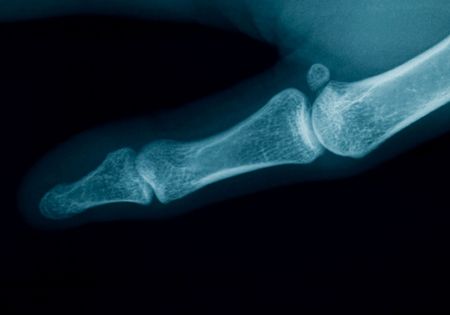
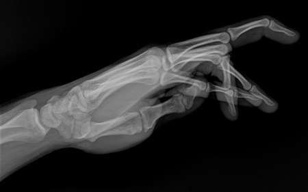
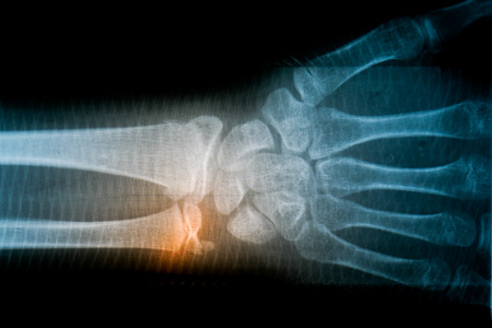
radiography of a middle aged woman finger
Коллекция по умолчанию
Коллекция по умолчанию
Создать новую

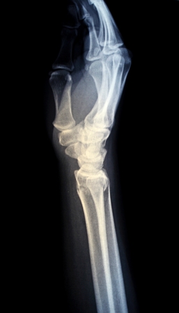
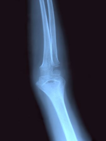
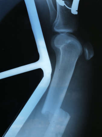
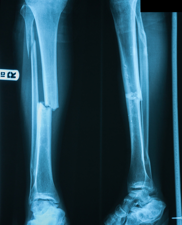
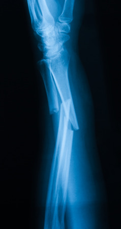
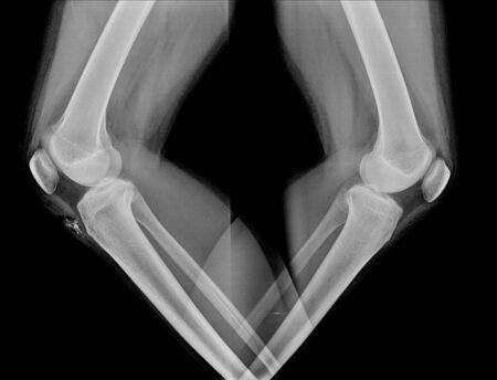
X-rays image of leg fracture patients
Коллекция по умолчанию
Коллекция по умолчанию
Создать новую
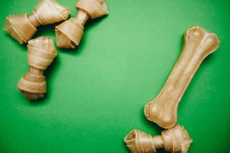
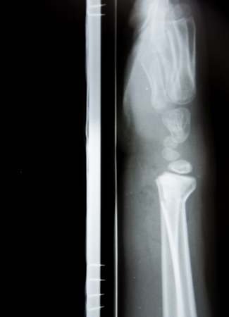
A snack bone and twisted dog treats on a bright one-color green background, close up, top view. Pet care and veterinary concept. Spase for your text or image.
Коллекция по умолчанию
Коллекция по умолчанию
Создать новую

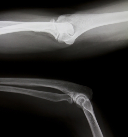
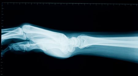
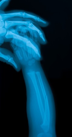
hand x-ray
Коллекция по умолчанию
Коллекция по умолчанию
Создать новую
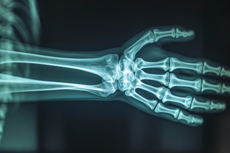
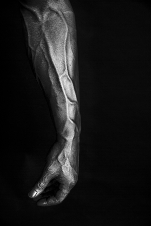
X-Ray image of human hands
Коллекция по умолчанию
Коллекция по умолчанию
Создать новую
Arm X RAY
Коллекция по умолчанию
Коллекция по умолчанию
Создать новую
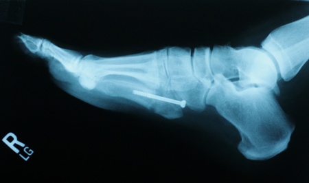
Jones Fracture
Коллекция по умолчанию
Коллекция по умолчанию
Создать новую
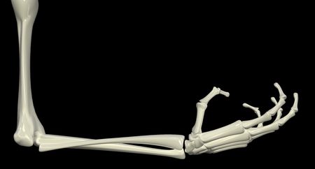
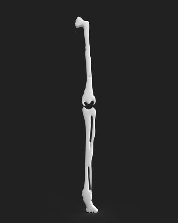
3d skeletal arm, isolated, dark background
Коллекция по умолчанию
Коллекция по умолчанию
Создать новую

X-ray of both human legs (broken legs)
Коллекция по умолчанию
Коллекция по умолчанию
Создать новую

X-ray medical picture - Human foot
Коллекция по умолчанию
Коллекция по умолчанию
Создать новую
Коллекция по умолчанию
Коллекция по умолчанию
Создать новую

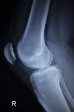
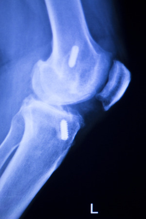
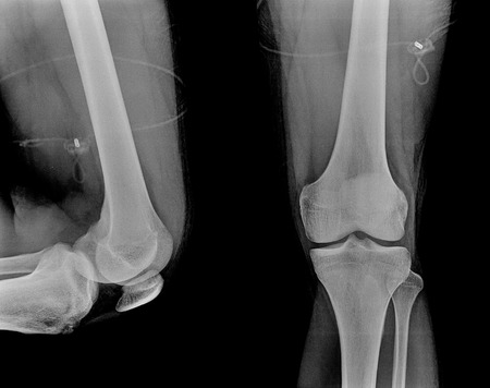
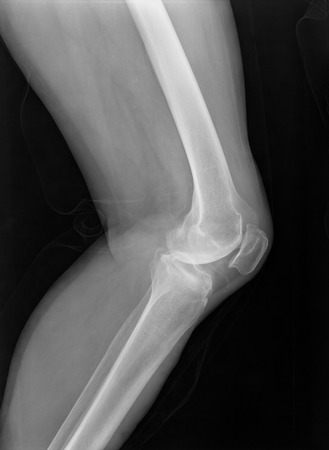
image of x-ray Osteoarthritis knee
Коллекция по умолчанию
Коллекция по умолчанию
Создать новую
Close up bone x-ray medical science background.
Коллекция по умолчанию
Коллекция по умолчанию
Создать новую

man's leg
Коллекция по умолчанию
Коллекция по умолчанию
Создать новую

knee with total replacement x-ray image on black background
Коллекция по умолчанию
Коллекция по умолчанию
Создать новую
This stock photo shows the X-ray of a fracture with displacement. High quality photo
Коллекция по умолчанию
Коллекция по умолчанию
Создать новую
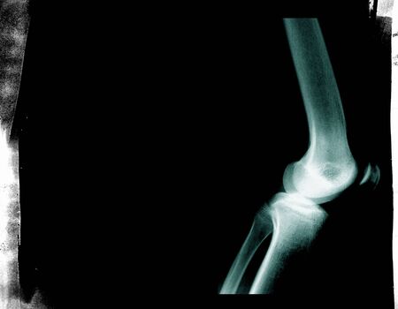
X-ray image of normal old age Knee
Коллекция по умолчанию
Коллекция по умолчанию
Создать новую
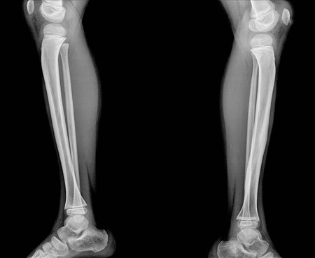
X-ray of both human legs (normal legs)
Коллекция по умолчанию
Коллекция по умолчанию
Создать новую
Bone anatomy skeleton body part human medical health care pain biology science arthritis x-ray injury leg disease white color orthopedic knee foot muscle physical person spine ache hospital femur
Коллекция по умолчанию
Коллекция по умолчанию
Создать новую
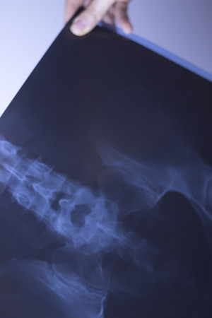
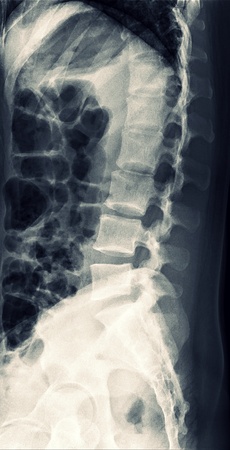
Medical hospital x-ray lowe back pain spine and hips traumatology scan.
Коллекция по умолчанию
Коллекция по умолчанию
Создать новую
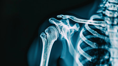
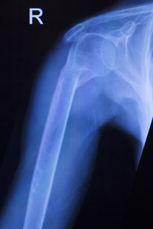
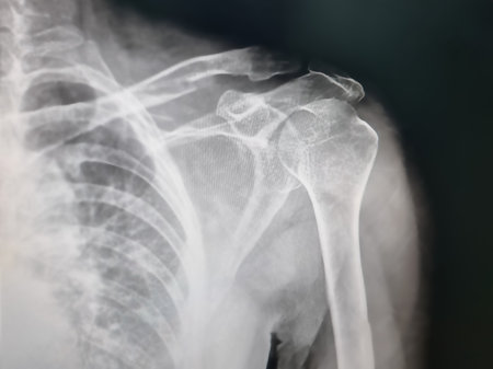
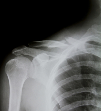
High-Resolution X-Ray Image of a Human Shoulder Joint. Concept of Medical Imaging, Human Anatomy, Diagnostic Tools, Orthopedic Study.
Коллекция по умолчанию
Коллекция по умолчанию
Создать новую
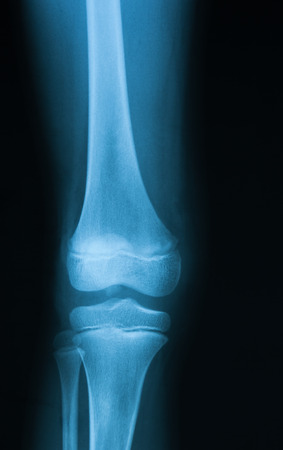
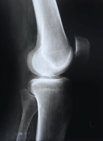
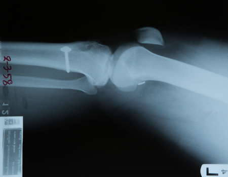
X-ray image of knee joint, AP view.
Коллекция по умолчанию
Коллекция по умолчанию
Создать новую
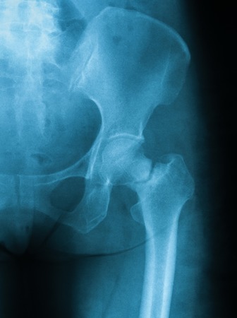
X-ray image of hip joint, AP view. Showing femural neck fracture
Коллекция по умолчанию
Коллекция по умолчанию
Создать новую
Xray of bent of human Hand fingers side view. X-ray of male hand and wrist. X ray of healthy whole bones of the hand close-up.
Коллекция по умолчанию
Коллекция по умолчанию
Создать новую

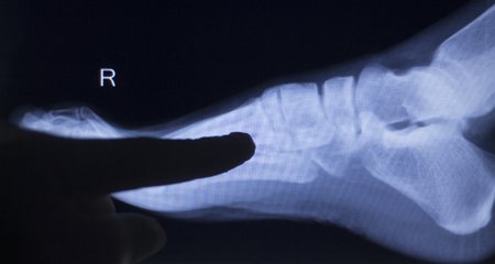
x-ray image of human foot joint , side view
Коллекция по умолчанию
Коллекция по умолчанию
Создать новую
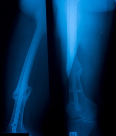
X-ray imag of leg, AP and lateral view, Show repeate fracture site after removed internal fixator.
Коллекция по умолчанию
Коллекция по умолчанию
Создать новую
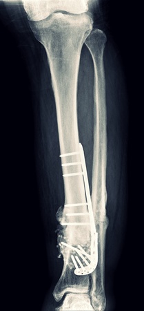
X ray fracture with fixation screws, broken leg
Коллекция по умолчанию
Коллекция по умолчанию
Создать новую

A pile of human bones in Chauchilla, an ancient cemetery in the desert of Nazca, Peru. The remains of many
people, some still with long hair, can be seen.
Коллекция по умолчанию
Коллекция по умолчанию
Создать новую

X-Ray Of Carpal And Metacarpal Bones In The Human Hand
Коллекция по умолчанию
Коллекция по умолчанию
Создать новую

White hairclips on purple background
Коллекция по умолчанию
Коллекция по умолчанию
Создать новую
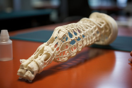
3d printer creating orthopedic splint, created with generative ai
Коллекция по умолчанию
Коллекция по умолчанию
Создать новую
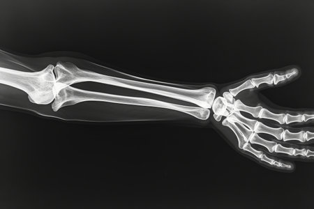
X-ray of human arm. X-ray image concept.
Коллекция по умолчанию
Коллекция по умолчанию
Создать новую
Shoulder joint injury xray traumatology and orthopedics test medical scan used to diagnose sports injuries in patient.
Коллекция по умолчанию
Коллекция по умолчанию
Создать новую
x-ray of the broken leg of beagle dog
Коллекция по умолчанию
Коллекция по умолчанию
Создать новую
Foot toes joint xray test scan results of patient with arthritis and joints pain in feet.
Коллекция по умолчанию
Коллекция по умолчанию
Создать новую

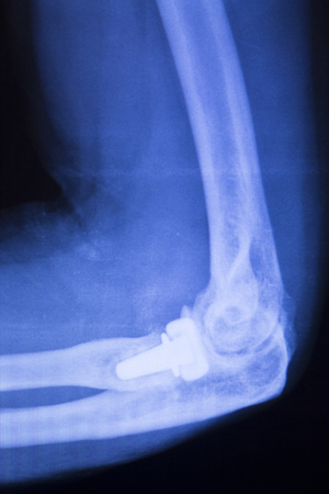
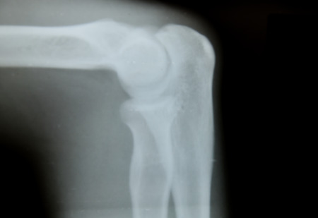
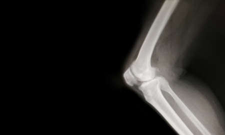
X-ray view of elbow
Коллекция по умолчанию
Коллекция по умолчанию
Создать новую

lower leg x-ray of a 48 year old female with a spiral fracture of the distal tibia
Коллекция по умолчанию
Коллекция по умолчанию
Создать новую
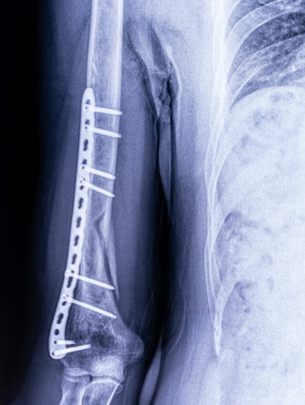
X-ray image of osteosynthesis of a humerus fracture in a man close-up
Коллекция по умолчанию
Коллекция по умолчанию
Создать новую
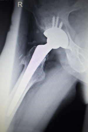
Hip joint replacement orthopedic titanium metal Traaumatology ball and socket implant x-ray image of old age patient.
Коллекция по умолчанию
Коллекция по умолчанию
Создать новую
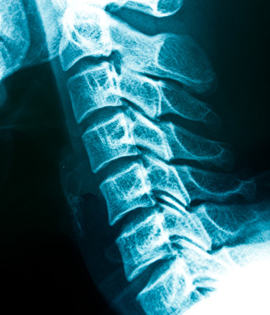
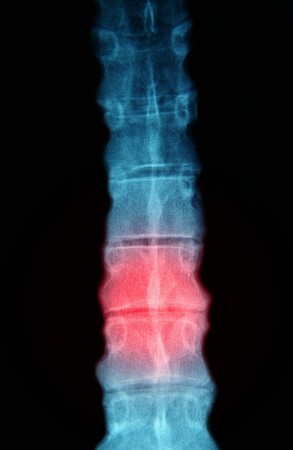
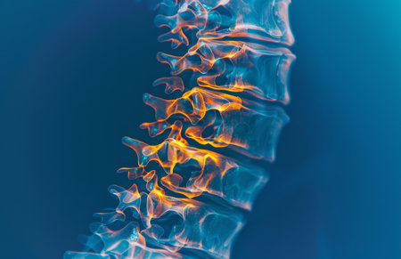
X-rays of the cervical spine
Коллекция по умолчанию
Коллекция по умолчанию
Создать новую
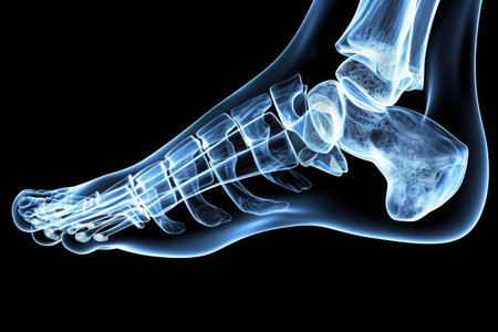
This x-ray image captures the intricate bone structure of a foot in vivid detail, Detailed 3D X-ray view of the human foot, AI Generated
Коллекция по умолчанию
Коллекция по умолчанию
Создать новую
x ray picture of wild animal skeleton
Коллекция по умолчанию
Коллекция по умолчанию
Создать новую
This x-ray image showcases a detailed view of a skeletons hand, capturing the structure and formation of the bones, A closer look at wrist bones through X-ray, AI Generated
Коллекция по умолчанию
Коллекция по умолчанию
Создать новую
Close up Thigh bone x-ray medical science background
Коллекция по умолчанию
Коллекция по умолчанию
Создать новую

X-rays image of leg fracture patients
Коллекция по умолчанию
Коллекция по умолчанию
Создать новую
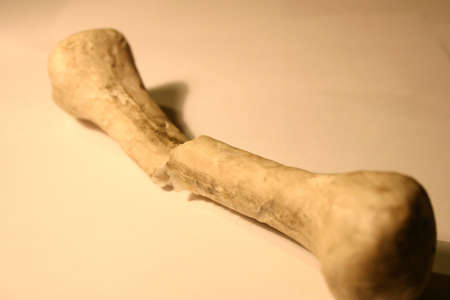
Bone
Коллекция по умолчанию
Коллекция по умолчанию
Создать новую
arm bone x-ray images blur and noise
Коллекция по умолчанию
Коллекция по умолчанию
Создать новую
X-ray orthopedic medical CAT scan of painful knee meniscus injury leg in traumatology hospital clinic.
Коллекция по умолчанию
Коллекция по умолчанию
Создать новую

snag
Коллекция по умолчанию
Коллекция по умолчанию
Создать новую

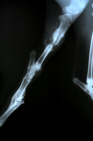
Dog broken limb radiography
Коллекция по умолчанию
Коллекция по умолчанию
Создать новую
X-ray of knees of both legs
Коллекция по умолчанию
Коллекция по умолчанию
Создать новую

X-ray Finger
Коллекция по умолчанию
Коллекция по умолчанию
Создать новую
This x-ray image captures a human hand, highlighting the presence of a bone, Detailed view of the ulna and radius bones in X-ray, AI Generated
Коллекция по умолчанию
Коллекция по умолчанию
Создать новую

Fractured Femur, Broken leg x-rays image
Коллекция по умолчанию
Коллекция по умолчанию
Создать новую
Inflammation, spine and 3D in xray for medicare, healthcare or radiology at hospital or clinic
Коллекция по умолчанию
Коллекция по умолчанию
Создать новую

x-ray bone malformation in femur, bone cancer
Коллекция по умолчанию
Коллекция по умолчанию
Создать новую

Side view of human knee-joint with kneecap on X-ray image
Коллекция по умолчанию
Коллекция по умолчанию
Создать новую

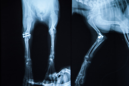
Implants and prostheses printed on 3D printer from biocompatible titanium powder. Plates for correction of deformation for animals. Titanium plates on animal bones printed on 3D printer.
Коллекция по умолчанию
Коллекция по умолчанию
Создать новую
X-ray of human shoulder (broken shoulder)
Коллекция по умолчанию
Коллекция по умолчанию
Создать новую
X-ray scan image of hip joints with orthopedic hip joint replacement implant head and screws in human skeleton in blue gray tones. Scanned in orthopedics traumatology surgery hospital clinic.
Коллекция по умолчанию
Коллекция по умолчанию
Создать новую
X-ray of knee
Коллекция по умолчанию
Коллекция по умолчанию
Создать новую
Knee joint implant screw xray showing in medical orthpodedic traumatology scan.
Коллекция по умолчанию
Коллекция по умолчанию
Создать новую

medical X ray image of knee
Коллекция по умолчанию
Коллекция по умолчанию
Создать новую
broken bone
Коллекция по умолчанию
Коллекция по умолчанию
Создать новую

Broken bone under the x-rays
Коллекция по умолчанию
Коллекция по умолчанию
Создать новую

Archaeological excavations and finds bones of a skeleton in a human burial , a detail of ancient research, prehistory.
Коллекция по умолчанию
Коллекция по умолчанию
Создать новую
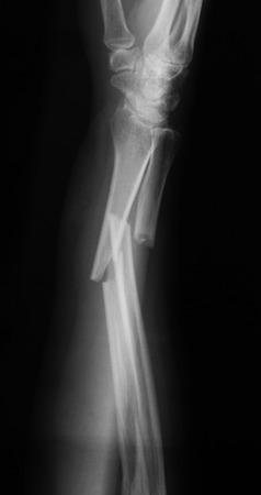
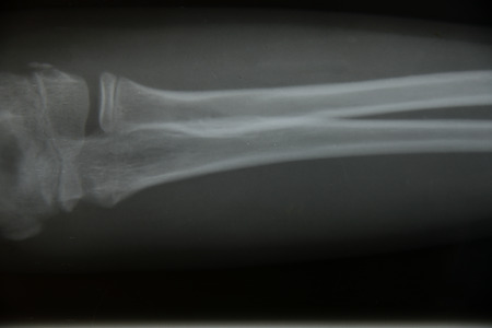
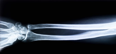
X-ray image of broken forearm, lateral view, show fracture of ulna and radius fracture.
Коллекция по умолчанию
Коллекция по умолчанию
Создать новую
X-ray Leg
Коллекция по умолчанию
Коллекция по умолчанию
Создать новую
X-Ray picture of knees front and side view
Коллекция по умолчанию
Коллекция по умолчанию
Создать новую
X-Ray Lumbar Spine
Коллекция по умолчанию
Коллекция по умолчанию
Создать новую

X-Ray photo of neck and skull.
Коллекция по умолчанию
Коллекция по умолчанию
Создать новую

Elbow joint tennis elbow inury x-ray test scan result before traumatology and orthopedics surgery.
Коллекция по умолчанию
Коллекция по умолчанию
Создать новую
knee bone computer x-ray images
Коллекция по умолчанию
Коллекция по умолчанию
Создать новую

arm splint, be in plaster cast
Коллекция по умолчанию
Коллекция по умолчанию
Создать новую

X ray of a human hand.
Коллекция по умолчанию
Коллекция по умолчанию
Создать новую

X-ray scan image of hip joints with orthopedic hip joint replacement implant head and screws in human skeleton in blue gray tones. Scanned in orthopedics traumatology surgery hospital clinic.
Коллекция по умолчанию
Коллекция по умолчанию
Создать новую

x-ray of an elbow dislocation
Коллекция по умолчанию
Коллекция по умолчанию
Создать новую
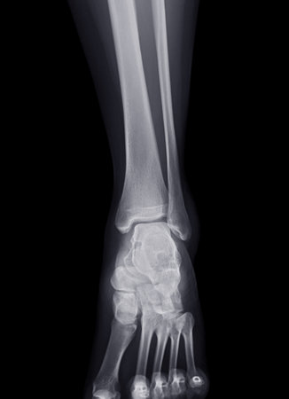
X-ray image of ankle joint for diagnosis fracture tibia and fibula bone.
Коллекция по умолчанию
Коллекция по умолчанию
Создать новую
x-ray image
Коллекция по умолчанию
Коллекция по умолчанию
Создать новую
X-ray image of broken forearm, lateral view, show fracture of ulna and radius
Коллекция по умолчанию
Коллекция по умолчанию
Создать новую
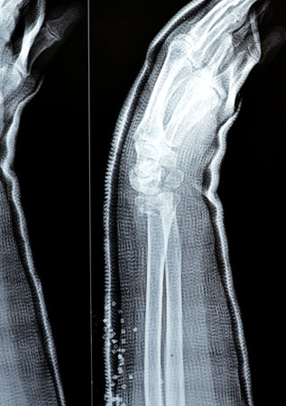
radiography of a middle aged woman hand and wrist
Коллекция по умолчанию
Коллекция по умолчанию
Создать новую

hand x-ray
Коллекция по умолчанию
Коллекция по умолчанию
Создать новую

x-ray of human leg (broken leg)
Коллекция по умолчанию
Коллекция по умолчанию
Создать новую
X-ray knee / Many others X-ray images in my portfolio
Коллекция по умолчанию
Коллекция по умолчанию
Создать новую
x-ray film skeleton human arm. health medical anatomy body concept
Коллекция по умолчанию
Коллекция по умолчанию
Создать новую
x-ray 2 knee joint ( Knee joint Lateral )on black background
Коллекция по умолчанию
Коллекция по умолчанию
Создать новую
X-ray image of a baby forearm with mother's hand.
Коллекция по умолчанию
Коллекция по умолчанию
Создать новую

x ray picture of wild animal skeleton
Коллекция по умолчанию
Коллекция по умолчанию
Создать новую

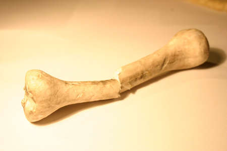
Black and white shot of a bone (from a pig). Vignetting was added.
Коллекция по умолчанию
Коллекция по умолчанию
Создать новую

x-ray of an injured elbow
Коллекция по умолчанию
Коллекция по умолчанию
Создать новую

Orthopedics knee joint meniscus, ligament, tendon and cartilage injury titanium modern metal implant X-ray scan.
Коллекция по умолчанию
Коллекция по умолчанию
Создать новую

X-ray Foot Ankle Calcaneus
Коллекция по умолчанию
Коллекция по умолчанию
Создать новую

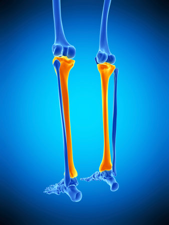
Polygonal vector illustration of leg bones on a dark blue background. Knee and ankle joints Polygon Design.
Коллекция по умолчанию
Коллекция по умолчанию
Создать новую
arm of muscle man
Коллекция по умолчанию
Коллекция по умолчанию
Создать новую
Colles fracture reduction of an old female, a type of fracture of the distal forearm, the broken end of the radius is bent backwards, as a result of a fall on an outstretched hand with osteoporosis
Коллекция по умолчанию
Коллекция по умолчанию
Создать новую
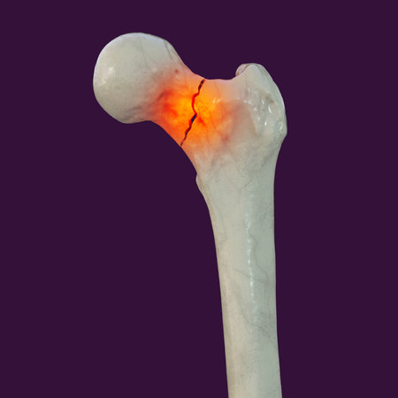
A fracture of the femur neck, a common type of hip fracture that typically occurs in older adults and can lead to mobility issues and other complications, isolated on white background, 3D illustration
Коллекция по умолчанию
Коллекция по умолчанию
Создать новую
medically accurate illustration of the tibia
Коллекция по умолчанию
Коллекция по умолчанию
Создать новую
abstract computer graphic background art wallpaper
Коллекция по умолчанию
Коллекция по умолчанию
Создать новую

close up finger x-ray film for check something
Коллекция по умолчанию
Коллекция по умолчанию
Создать новую

X-Ray image of human hand a fracture on the metal support as medical examination
Коллекция по умолчанию
Коллекция по умолчанию
Создать новую
Legion-Media
Создайте свои проекты на основе качественных стоковых фотографий и видео.


Copyright © Legion-Media.